

In short
- A beauty mark and a mole are the same lesion: a melanocytic nevus, a benign growth of pigment cells1.
- First the lesion is examined, then a method is chosen. The examination usually includes visual inspection and dermoscopy, and checks for warning signs23.
- Excision and shave removal leave a sample for pathology; laser and cautery do not, which is why they are not used to remove a pigmented mole34.
- No removal is completely markless. Every method may leave a scar or a change in color, to a varying degree56.
- A mole that is removed is usually sent for pathology, because a small proportion of lesions that look benign turn out otherwise3.
- At the clinic, the removal is performed by Dr. Dana Buchbut herself, a physician (M.D.).
What is the difference between a beauty mark and a mole?
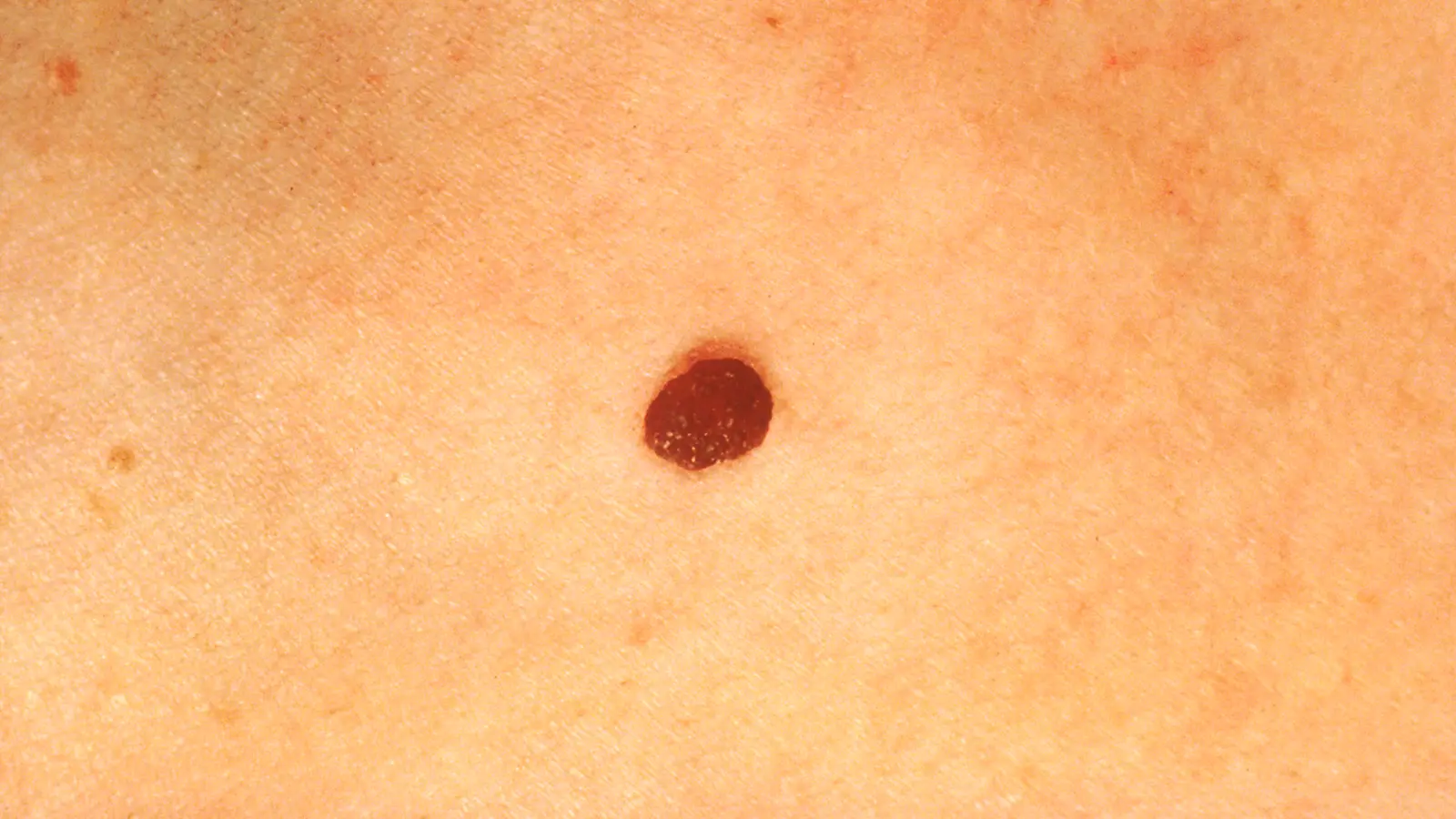
There is no difference: a beauty mark and a mole are two everyday names for the same lesion, a melanocytic nevus, a benign, localized growth of the pigment cells in the skin. The number of moles a person has depends on genetics, sun exposure and their immune system, and moles can appear and change in appearance over the course of life, mainly in childhood and adolescence1. Part of this page deals with ordinary moles that have already been diagnosed as benign; if the question is how to recognize a mole that needs an urgent examination, there is a separate page for that, and a full explanation of the types of skin lesions can be found on the main page on skin lesions.
When is a mole removed: for medical or cosmetic reasons?
There are two main reasons to remove a mole: a clinical suspicion that needs investigation, or an aesthetic wish to remove a known benign lesion. In most cases moles are a normal, benign finding that does not need to be touched at all. Removal for a medical reason is considered when there are warning signs on examination, when the lesion changes, itches or bleeds, or when it is located where it is constantly injured, for example by rubbing against clothing71. Purely aesthetic removal is possible once the lesion has already been diagnosed as completely benign. In both cases the order of steps is the same: first establish what the lesion is, and only then choose how to remove it. Other benign lesions that are usually removed for aesthetic reasons, such as skin tags (acrochordons), are described on a separate page. What the health fund (kupat holim) covers when removal is medical, and what applies when it is aesthetic, is explained on the page mole removal through the health fund.
What is checked before removal?
Before removal is recommended, the lesion is examined clinically, usually with dermoscopy, to make sure it has no warning signs. Dermoscopy is an examination with an optical device that magnifies and illuminates the skin, making it possible to see structures inside the lesion that are not visible to the naked eye. The examination applies the ABCDE rule (asymmetry, border, color, diameter and evolution) and asks whether the lesion is new, whether it has changed, and how it looks compared with the person's other moles2. Only after the clinical picture looks benign do you move on to the next step: choosing the removal method, which itself depends on whether a sample needs to be kept for further testing38.
Mole removal methods at a glance
| Method | How it is done | Sample for pathology | What is important to know |
|---|---|---|---|
| Excision with stitches | An elliptical cut, with healthy margins around the lesion | Yes, the entire lesion | The most complete removal and the lowest risk of recurrence, but a linear scar remains |
| Shave removal | Horizontal removal with a blade, without stitches | Yes | Usually a better cosmetic result, but a higher risk that the lesion comes back |
| Laser, plasma or cautery | Destruction of the tissue in place | No | Not recommended for a pigmented mole; suitable for some other lesions, and only after diagnosis |
Healing usually takes two to three weeks, and a removed mole is usually sent for pathology testing.
How is a mole removed by excision, and when is it the right choice?
Elliptical excision with stitches removes the entire lesion, including healthy margins around it, and is considered the "gold standard" for completeness of removal, but it is also the method that leaves the most noticeable linear scar. A meta-analysis that reviewed 46 studies and 4,201 lesions found that excision achieved complete removal in 96.4% of cases and an especially low recurrence rate of only about 2.1%. In the same meta-analysis, however, excision received the lowest cosmetic satisfaction score of the methods examined, about 6.2 out of 109. Excision is particularly suitable for flat lesions, deep lesions, and any case in which it is especially important to ensure complete removal and a complete sample for pathology.
What is shave removal, and what is the risk that the mole comes back?
Shave removal takes off the lesion with a horizontal motion of a blade, without stitches, and usually gives a better cosmetic result, but with a higher risk that the lesion comes back. A review on the management of moles reported a recurrence rate of 13% to 26% after shave removal3. In a randomized trial comparing shave removal with elliptical excision, the recurrence rate after shave removal alone was 11.7%, and about 94% of recurrences appeared within three months; at the same time, shave removal had more involved margins on pathology, but a higher cosmetic result and satisfaction compared with excision10. Another study, on intraoperative monitoring, found a recurrence rate of about 33%, and in 5% of cases not enough tissue remained to make a definite diagnosis from the sample11. The main drawback of shave removal is that the procedure is relatively "blind" compared with excision, because the physician does not see the full depth of the lesion during removal3.
Can a mole be removed with laser or plasma?
Not a pigmented lesion. Laser, plasma ablation and cautery vaporize or destroy the tissue in place, so no sample remains that can be sent for laboratory testing36. The official guideline of the German Dermatological Society (S2k, 2022) states explicitly that "laser treatment of pigmented melanocytic nevi is not recommended"4. The position paper of the European Society for Laser Dermatology adds that in all cases "accurate identification of the disorder is mandatory" before choosing laser treatment for a skin lesion8. Cleveland Clinic also writes that laser, cautery and cryotherapy are "usually not recommended" for mole removal6. The practical reason: if the lesion was not the harmless mole it appeared to be, but an early stage of melanoma, the diagnosis may be lost permanently after laser treatment. Laser may be suitable for other, non-melanocytic lesions, such as skin tags, seborrheic keratosis or xanthelasma, but there too only after diagnosis. Laser hair removal in an area with moles can also change the appearance of the moles, so it is worth pointing them out before such treatment12.
In short: which method leaves a sample for pathology? This is the question worth asking before any removal, including when laser removal is offered.
| Method | Is tissue left for examination? | Mainly suitable for |
|---|---|---|
| Excision (with stitches) | Yes, the whole lesion | A flat mole, a mole that has changed, or when it matters to remove the entire lesion |
| Shave removal | Yes, the raised part | A raised mole that looks benign |
| Laser (CO2, erbium and others) | No, the tissue is vaporized | Lesions that are not moles, after a clear diagnosis, for example skin tags, seborrheic keratosis or xanthelasma |
| Cautery or plasma | No | Skin tags and superficial non-pigmented lesions, after diagnosis |
When you are offered laser removal of a mole, it is worth asking three questions: what is the diagnosis, will a sample remain for pathology, and what happens if the mole comes back. For flat moles, which are usually removed by excision, see flat mole, and for what makes up the cost of each method see how much mole removal costs.
Can a mole on the face be removed without leaving a mark?
No. No removal method promises a result with no mark at all, and the face is especially sensitive because every small change is more noticeable. The American Academy of Dermatology writes that "any time you injure your skin, there is a risk of scarring"5, and Cleveland Clinic notes that after mole removal a scar remains that "may or may not be noticeable"6. The data support this: even with the methods that had the highest cosmetic scores in studies, Er:YAG and CO2 lasers, with scores of 8.8 and 8.4 out of 10, a result was still measured, not a complete absence of a mark9. The realistic goal is as small a mark as possible, and how noticeable it is depends on the method, the location of the lesion on the face, its depth and the skin type3910. On removal from the nose, lip and eyebrow, see mole removal on the face, and on what is normal for the scar that remains, scar after mole removal.
What do you do after mole removal, and how long does healing take?
Keep the wound clean and moist, change the bandage every day, and once it has closed, protect it from the sun. Healing usually takes two to three weeks. According to Cleveland Clinic, after removal petroleum jelly is applied to the area and covered with a bandage, which is cleaned and changed daily. You can return to work or school the same day, and mild stinging in the first days is expected6.
Why moist and not dry: the American Academy of Dermatology explains that petroleum jelly prevents the wound from drying out and forming a scab, and that a wound with a scab heals more slowly. Wash gently with water and mild soap. After the wound has healed, apply broad-spectrum sunscreen (SPF 30 or higher), as this may reduce redness or darkening and help the scar fade faster13. If there were stitches, the time for removing them is set at the clinic according to the location of the incision.
See a doctor if the bleeding does not stop, if spreading redness or pus appears, or if the mole starts to grow back in the same place6. On treating a scar that remains noticeable: scars.
Why is the mole sent for pathology?
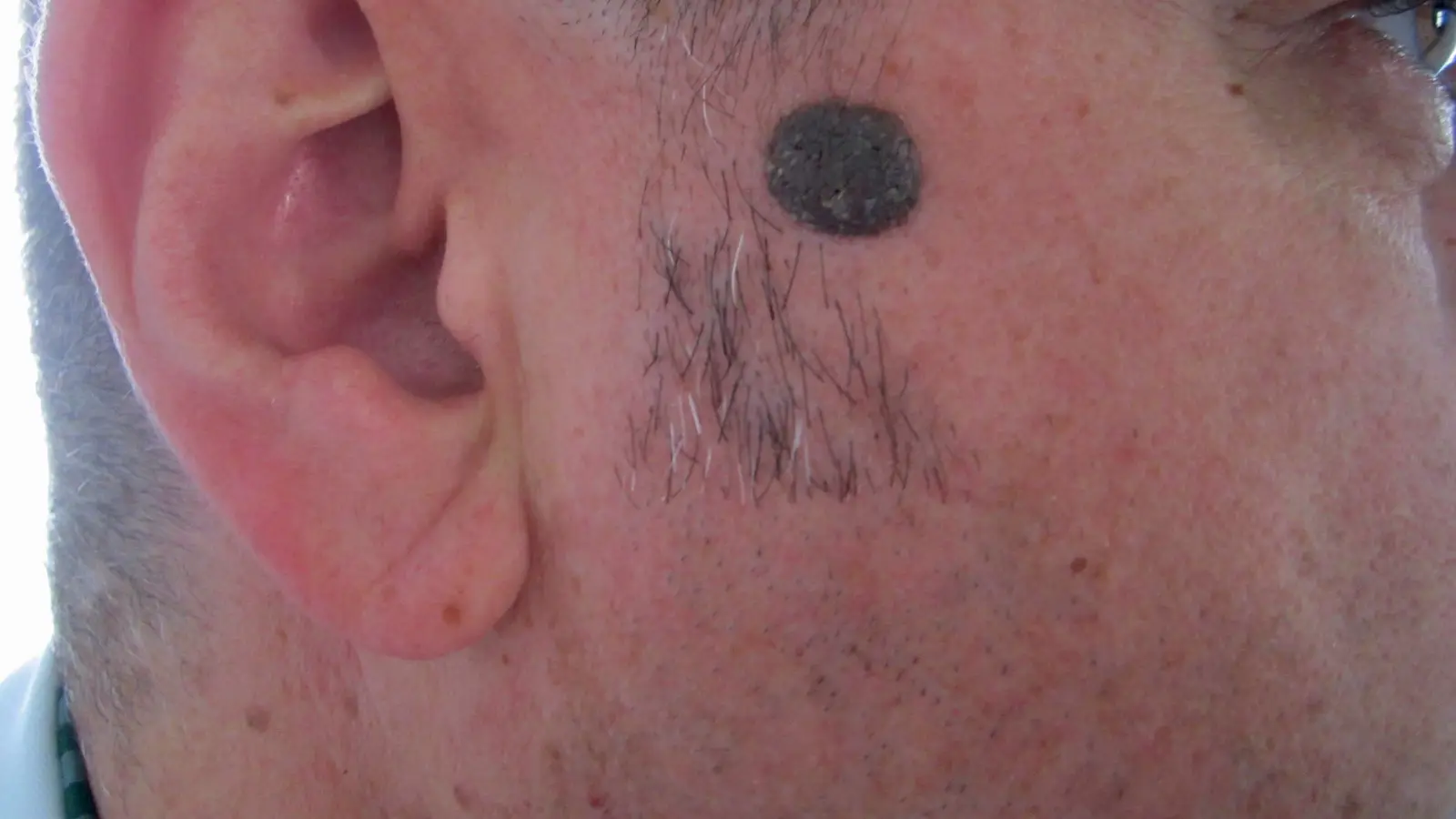
Because even a lesion that looks completely benign to the eye can turn out differently under the microscope, and this is the only way to know for certain. A review on the management of moles states that the accepted practice is to send every removed nevus for histological examination, and according to a study cited in it, 2.3% of nevi diagnosed clinically as benign turned out under the microscope to be malignant tumors: melanoma, basal cell carcinoma or squamous cell carcinoma3. Another study, which examined about 1,815 lesions excised on the assumption that they were benign, found that 0.33% of all lesions were actually malignant, and recommends routine pathology mainly for solid, pigmented or ulcerated lesions14. The American Academy of Dermatology also notes that a removed mole is examined under the microscope for cancer cells5. Methods that remove tissue, excision and shave removal, allow this examination; methods that destroy the tissue in place, such as laser, do not. How to read the report that comes back from the laboratory is explained on the page the pathology report after mole removal. When comparing price quotes, make sure this examination is included; see how much mole removal costs.
What happens if the mole comes back?
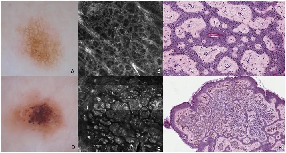
A mole that comes back after partial removal needs attention, because it can look like melanoma, both clinically and under the microscope. The phenomenon is sometimes called a "recurrent nevus" or, mistakenly, "pseudomelanoma," and it results from the removal method, usually shave removal or another partial method, not taking out all the pigment cells3. A study that examined the histological and immunohistochemical findings in recurrent lesions found that they can mimic melanoma under the microscope, which complicates diagnosis15. The practical meaning: a new lesion that appears exactly where a mole was removed in the past is a reason to report it to a doctor, not to assume it is a routine, meaningless phenomenon.
Why not remove a mole at home?
Because home mole-removal products can cause permanent damage, and even when they "succeed" in removing the lesion, they skip the diagnosis step, exactly the part that protects you. The US FDA has documented serious skin injuries, including burns, ulcers and permanent scars, caused by unapproved products for removing moles and skin tags; a considerable share of the cases were on the face, close to the eyes16. Beyond the direct risk to the skin, self-removal gives up the examination that confirms in advance that the lesion is benign, and the option of sending a sample to pathology when needed. What these products contain, and what to do if you have already tried them, is on the page removing moles at home: the risks.
How is it done at Dr. Dana's clinic?
Dr. Dana Buchbut examines every mole herself before she recommends a removal method. Only after the clinical picture looks benign do you move on to the procedure, so that you know exactly what to expect even before booking an appointment. A mole that is removed is sent for pathology testing, so removal is done with methods that leave a sample for testing. For a mole that looks suspicious, or that has changed, the recommendation is to be examined first by a dermatologist, as detailed on the page on suspicious moles. An overview of the types of lesions and removal methods can be found on the page on skin lesions.
What is new in research (updated November 2026)
Studies published in 2024–2026 that were not yet covered on this page, in brief. They are presented to reflect the current state of knowledge, not as a promise of results. A decision about treatment is always made after a personal examination.
- Dermatoscope-guided shave removal (2024): a study of 64 patients with facial moles in dark skin examined layered shave removal, guided by a dermatoscope until clear tissue was seen, followed by cautery of the base. In most patients the cosmetic result was good or fair, and recurrence of the mole was uncommon. The study had no comparison group.17
- Artificial intelligence in mole examination (2026): a meta-analysis of prospective studies in the journal JAMA Dermatology found that artificial intelligence systems that analyze dermoscopic images reach accuracy similar to that of dermatologists in diagnosing melanoma, and may improve accuracy as an aid to the physician. Most of the studies were at risk of bias, so these tools do not replace a medical examination and pathology testing.18
- Laser for pigmented lesions (2025): a review of 77 studies on lasers for benign pigmented lesions (such as café-au-lait spots and nevus of Ota) found that recurrence and color changes after treatment are common, especially in dark skin. The review does not deal with ordinary moles, and for them the recommendation above still stands: prefer a method that allows pathology testing.19
Terms on this page: Dermoscopy · Pathology testing · Skin tag · Liquid nitrogen freezing · Seborrheic keratosis · Full glossary
Frequently asked questions
Is mole removal painful?
Most removal methods are performed under local anesthesia, so the procedure itself should not hurt. After the anesthesia wears off you may feel mild tenderness in the area, which usually goes away on its own.
How long does it take to heal?
It depends on the method and the size of the lesion. A stitched wound or a small open wound needs a few days to weeks for the skin to close, and the final appearance of the area keeps changing and improving for months after the procedure, as happens with any scar.
How do you care for the wound after mole removal?
Wash gently with water and mild soap, apply petroleum jelly so the wound stays moist and does not form a scab, and change the bandage every day. After the wound has closed, apply sunscreen to it. See a doctor if there is bleeding that does not stop, spreading redness or pus.
Can the mole come back?
Yes, mainly after methods that leave some of the cells in place, such as shave removal or cautery. If something reappears in the area where a mole was removed, that is a sign to report it to a doctor and not to assume it is routine.
Are stitches needed?
It depends on the method: an elliptical excision is usually closed with stitches, whereas shave removal leaves a flatter wound that usually heals without stitching. The choice between the methods is made according to the location, size and depth of the lesion.
When do you get the pathology result?
The sample is sent to the laboratory and the result usually arrives within a few weeks, depending on the laboratory's workload. The clinic that performs the removal is the one that gets in touch when the result arrives.
Can a flat mole be removed?
A completely flat mole is less suitable for shave removal, because shaving requires "catching" tissue that protrudes above the skin surface. Excision, which also removes the deeper layers of the lesion, is more suitable for a flat lesion.
Why are moles not removed with laser?
Because laser vaporizes the tissue in place and leaves nothing of it for laboratory testing. The official guideline of the German Dermatological Society states that laser treatment of pigmented lesions such as moles is not recommended, for exactly this reason.
Can laser hair removal harm moles?
A systematic review found that laser hair removal can change the clinical and dermoscopic appearance of moles in the treatment area, to the point of resembling an atypical lesion. It is worth pointing out which moles are in the area before laser hair removal.
Is it okay to remove a mole yourself, at home?
No. Regulatory bodies have documented serious skin injuries (burns, ulcers and permanent scars) from unapproved mole-removal products sold for self-use. Beyond the direct risk, self-removal skips the diagnosis step, which is exactly the part that protects you.
Related reading
Skin lesions
The main guide: which lesion you have, and how it is identified before removal.
Read (Hebrew)Suspicious mole
ABCDE and warning signs: when a mole needs an urgent examination.
Read (Hebrew)Scar treatment
If a noticeable scar remains after removal, it has evidence-based treatments of its own.
Read (Hebrew)Facial capillaries
Red capillaries on the nose, spider veins and red spots on the skin: what causes them (rosacea, sun, pregnancy, liver), when they need investigation, and what treatments exist. Medical information with sources.
Read (Hebrew)Lipoma
A lipoma is a soft, benign lump of fat under the skin. How it is diagnosed, which signs require investigation, which removal methods exist, the chance of recurrence and what healing looks like.
Read (Hebrew)Milia
Milia, hard white bumps on the face and under the eye: what causes them, why not to squeeze them at home, when they go away on their own and which removal methods exist, including risks.
Read (Hebrew)Abscess and furuncle
An abscess or furuncle (boil) is a pus-filled skin infection. What you can do at home, why not to squeeze it, when drainage by a doctor is necessary, when antibiotics are added and which signs require urgent care.
Read (Hebrew)Mole removal on the face
What is different on the nose, lip, eyebrow and cheek, and how to choose a method that leaves a small mark.
Read (Hebrew)Scar after mole removal
What is normal in the first weeks, what helps and when the scar is treated.
Read (Hebrew)Pathology report after mole removal
What the terms in the report mean and what to do next.
Read (Hebrew)Raised mole
Why a mole is raised, when to have it checked and how it is removed.
Read (Hebrew)Mole on the scalp
When to have it checked, and how it is removed without harming the hair.
Read (Hebrew)Wound care after lesion removal
Dressings, showering, sun and when to come back for a check.
Read (Hebrew)Skin lesion removal in Tel Aviv
What is checked before removal, which lesions are removed, pathology and healing, at the clinic in Ramat HaHayal.
Read (Hebrew)Flat mole
How to tell it from a sunspot, when to have it checked, and why it is removed by excision rather than shaving.
Read (Hebrew)Mole from birth (brown birthmark)
Congenital nevus in adults: types by size, what changes, when it is removed and when to refer.
Read (Hebrew)Removing moles at home: the risks
Creams, black salve, plasma pens and thread: what they do to the skin, and what to do if you have already tried them.
Read (Hebrew)How much mole removal costs
What determines the price, what to ask in a quote and why removal without pathology is a false saving.
Read (Hebrew)Mole removal through the health fund
When the health fund (kupat holim) covers it, what the pathway looks like, and what to ask your supplementary insurance.
Read (Hebrew)The information on this page is general and for education only. It does not replace an examination, diagnosis or personal medical advice. A decision about treatment is made only after an examination.